
Brief Summary
Toshimasa Kumazaki, Tomihisa Takahashi, Takashi Nakano, Tetsuo Sakai
https://www.jstage.jst.go.jp/article/jmj/68/4/68_JMJ22-0009-OA/_article/-char/ja/
In this month's research summary we turn our attention to the hip flexors as we review a paper by Kumazaki et al, looking into the difference in action between the iliopsoas and rectus femoris muscles in hip flexion. It has been widely reported that the hip flexors play a crucial role in maximal sprinting, subsequent biomechanical analysis employing EMG identified a combination of iliopsoas and rectus femoris contribution. We also know that muscle fiber length (FL) and physiological cross-sectional area (PCSA), are well known to affect the muscle function, including the contraction speed, maximum muscle strength, and the effective contractible range. This study aimed to quantify the iliopsoas and rectus femoris contributions to maximal hip flexion torque using PCSA and moment arm length (MAL) in the sagittal plane. Morphometric data from skeletal specimens were used to estimate individual muscle flexion speed by calculating flexion from 1% muscle shortening. While these methods did not measure actual joint torque and speed, they theoretically revealed the maximal and relative muscle contributions.
Method
Twenty-two lower limbs from 10 male and 12 female adult Japanese cadavers, all without apparent hip or knee joint degeneration were used for analysis. For morphometry and PCSA estimation of the iliopsoas and rectus femoris, the author used 12 specimens from 6 male and 6 female cadavers (mean age 78.6 ± 10.6 years). For MAL measurement, 10 specimens from 4 male and 6 female cadavers (mean age 79.6 ± 6.8 years) were used. In the isolated muscle specimens, the mass (M), FL, and pennation angle (θ) were measured. Based on these data, the PCSA was calculated using the following formula, PCSA (cm2) = M (g) × cos θ/ρ (g/cm3) × FL (cm)
The MAL in the hip extension position was calculated from three-dimensional reference values of reflective markers attached to the skeleton that were measured with four infrared cameras and an optical motion capture system. To estimate the relative contributions of individual flexors to maximal hip flexion torque, the authors calculated the product of PCSA and MAL for the psoas major (PM), iliascus (IL), and rectus femoris. These indicators of maximal hip flexion torque were determined for each muscle. The relative contributions were expressed as ratios of these indicators to the total hip flexion torque for the three muscles across various hip joint angles from 0° (extension) to 90° (flexion).
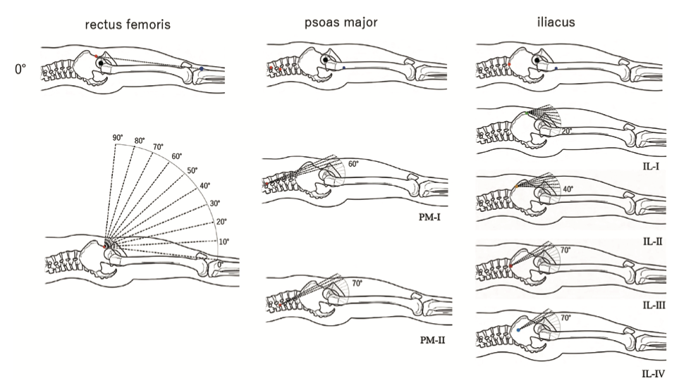
Figure 1. Measurement of moment arm length (MAL) of the three muscles under different hip joint angles.
Result
The authors found that In hip extension (0°), the MAL of the rectus femoris (35.0 ± 4.0 mm) exceeded that of the PM and IL (22.9 ± 1.9 mm). As the hip flexed, the rectus femoris MAL increased, peaking at 40° (50.4 mm), then decreased up to 90°. The PM-I MAL was slightly larger than PM-II between 60° and 90°, with similar changes during flexion. The IL-I and IL-II MALs remained constant up to 10° and 30°, then increased steeply up to 90°, surpassing the rectus femoris MAL between 40° and 60°. The IL-III and IL-IV MALs remained constant up to 50° and 60°, then increased steeply up to 90°, surpassing the rectus femoris MAL between 70° and 80°.
At 0° hip extension, the rectus femoris contributed 57.3%, increasing to 60.4%-62.3% at 20°-40°, then decreasing sharply in deep flexions, falling behind the iliopsoas at 60°-70°. The iliopsoas contribution remained steady up to 40°, then increased gradually, surpassing the rectus femoris at 80°-90°. The PM contribution gradually decreased up to 50°, then increased in deep flexions, matching the rectus femoris at 90° (Figure 2).
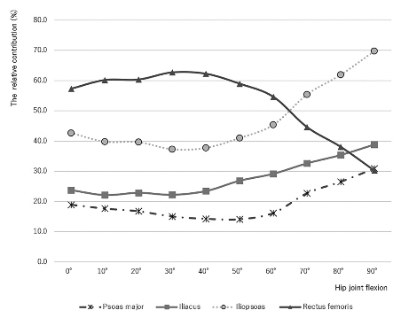
Figure 2. Relative contribution during maximal hip flexion torque.
At 0° hip extension, the PM and IL rotated 2.6 and 2.1 times faster than the rectus femoris, respectively. The PM rotation speed remained constant up to 50° flexion, then decreased steeply to 90°. The IL rotation speed stayed constant up to 10° flexion, then decreased gradually to 90°. The rectus femoris rotation speed decreased slightly up to 40°, reaching about 90% of its speed at 0°, then increased slightly up to 90°, returning to its initial speed.
This study identified two key functional parameters for the main agonists of hip joint flexion: the relative contribution to maximal flexion torque and the relative rotation speed during muscle contraction. These parameters highlighted distinct functional properties of the iliopsoas and rectus femoris. These findings have significant implications on exercise prescription for developing specific strength adaptations and rehabilitation programs. Findings can be applied to KT360 when selecting appropriate hip flexion screening/monitoring and training protocols. If practitioners are aiming for a protocol with contribution biased towards iliopsoas then Hip Flexion 90 should be selected, alternatively if we look to bias towards rectus femoris then Hip Flexion 60 or 20 should be the desired protocols.
For more information on Hip Flexion protocols on KT360 for screening / monitoring or training purposes please contact support@kangatech.com.